

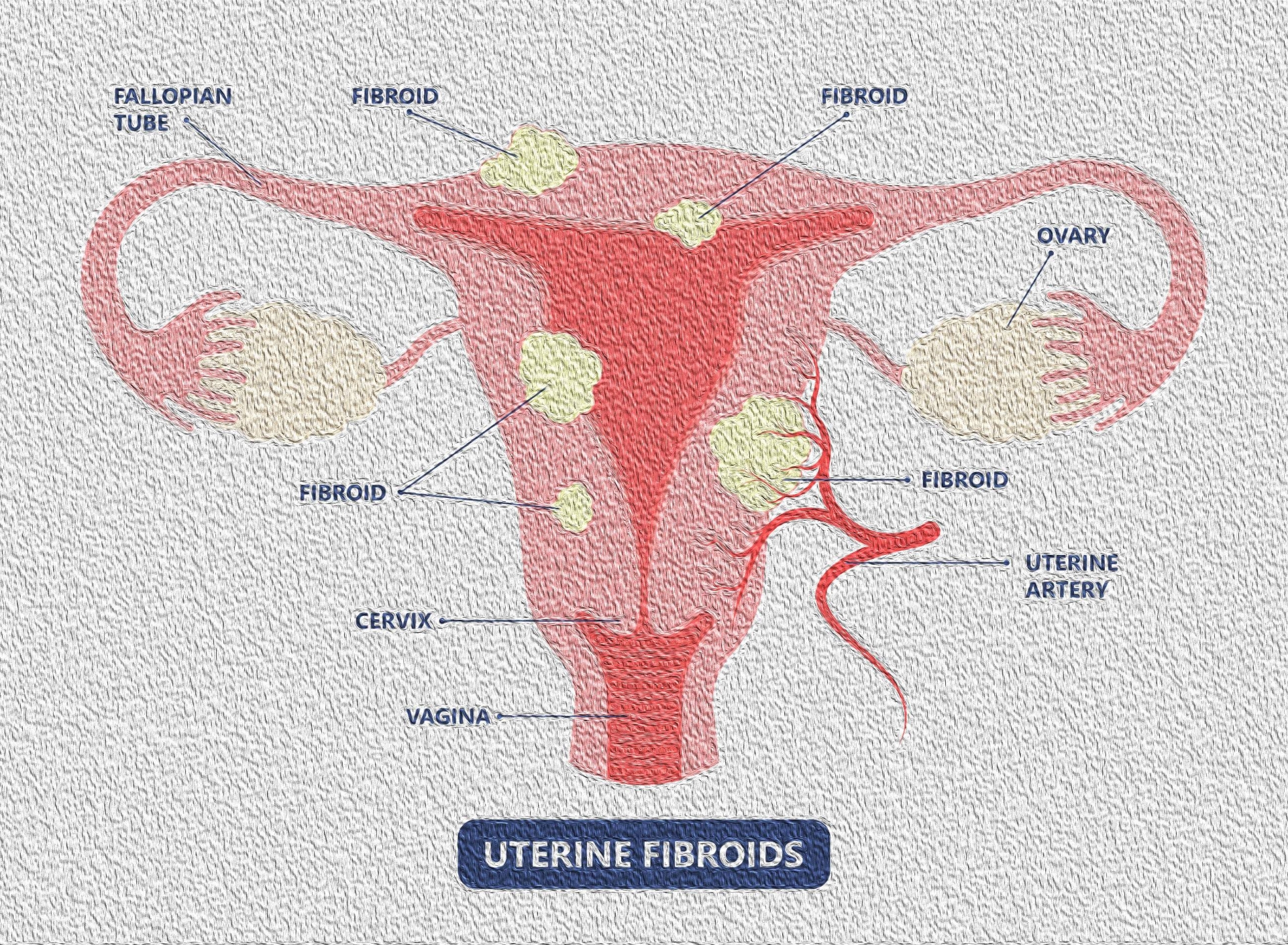
For a successful surgical outcome it is important to identify preoperatively the number, size, location and depth of intramural extension of the uterine myoma. These are determinants of complete resectability, the number of surgical procedures necessary and the duration of surgery.
Hysteroscopic classification of myomas
The European Society of Hysteroscopy classification system is based on myoma location and the amount of myoma mass protruding or encroaching on the endometrial cavity. Type 0 myomas are pedunculated with the myoma lying completely within the endometrial cavity. Type I myomas are described as sessile with less than 50% intramural extension. Finally, type II myomas are submucosal in location, with more than 50% intramural extension.
The degree of surgical difficulty is related to the depth of penetration and size of the myomas.
Pedunculated Type 0 myomas up to 3 cm on diameter can usually be easily removed hysteroscopically. Larger (>3cm) Type 0 and Type I myomas can be approached hysteroscopically, with careful monitoring of operating time and balance of fluids, due to the increased fluid intravasation via myometrial venous channels during resection. Often it is safer and thus preferable to remove larger myomas by two separate procedures. Only the most experienced hysteroscopists should attempt a hysteroscopic resection of a Type I myomas of 5 cm or larger.
Hysteroscopic resection of Type II myomas should only be approached by the most skilled hysteroscopists and after considering also of an alternative approach via laparoscopy or laparotomy. This is because hysteroscopic resection of Type II myomas results in extensive damage to the endometrium, resulting in an increased risk of Asherman’s syndrome as well as decreased fertility and hypomenorrhea. For these reasons patients are often treated postoperatively with estrogens and progestins, intrauterine stents and balloons or “second look” hysteroscopy. Because of this risk, hysteroscopic removal of Type II myomas should be performed with exceptional caution in women who desire future fertility.

