

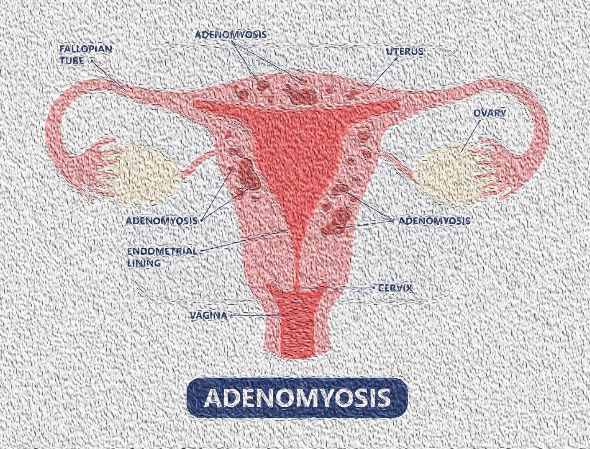
Patients with adenomyosis will usually report symptoms like those reported by patients with endometriosis. Common complaints include menorrhagia, dysmenorrhea, metrorrhagia, chronic pelvic pain and dyspareunia.
Currently, adenomyosis remains a largely clinical diagnosis as definitive diagnosis of adenomyosis requires a histologic exam of uterine tissue. Typically, this step is circumvented in women desiring future fertility or who wish to preserve their uterus. However, increasing availability and technological advances in imaging such as ultrasound, CT and MRI scans have improved evaluation for adenomyosis, but it remains largely under-diagnosed until the time of hysterectomy. Therefore, diagnostic hysteroscopy with myometrial biopsyand endometrial biopsy has been found to be indispensable in identifying suspected adenomyosis.
Hysteroscopy can definitively prove or exclude the presence of adenomyosis due to its direct visualization, while examination and biopsy of the endometrial surface can demonstrate features which would support adenomyosis. These may include:
- Irregular endometrium with superficial openings
- Irregular sub -endometrial myometrium (junctional zone) on histologic exam
- Irregular myometrial architecture on histologic exam
- Intramural endometriomas
Myometrial biopsy has long been considered as a possible means for obtaining a specimen for histologic diagnosis without requiring hysterectomy. Myometrial core needle biopsy can be performed under direct visualization during diagnostic hysteroscopy for the evaluation for chronic pelvic pain or infertility. Core needle biopsy obtained under direct visualization from an area that has been evaluated with ultrasound and suspected to have adenomyosis, has a sensitivity of 98% and a specificity of 100%.
Alternatively, a transvaginal approach for myometrial core needle biopsy under ultrasound guidance is feasible by a trained physician.
The only definitive treatment for adenomyosis is hysterectomy; however, this is not an option for patients who desire future fertility and may not be an option for patients who are poor surgical candidates. The medical management consists of anti-inflammatory medications and hormonal therapies. Hormonal therapies cause ovarian suppression, mainly through negative feedback on the hypothalamic-pituitary-ovarian axis. By suppressing ovarian function, hormonal stimulation of adenomyotic tissue is suppressed.
Conservative management of adenomyosis consists of Uterine Artery Embolization (UAE), Adenomyomectomy and most recently High Intensity Focused Ultrasound (HIFU).
HIFU is a conservative surgical method which allows patients to preserve their uterus. MRI or ultrasound imaging is used to visualize the uterus and direct high-intensity ultrasound beams at a targeted area within the myometrial tissue. These targeted ultrasound beams cause thermal ablation and necrosis. HIFU can be used on both focal and diffuse adenomyosis. Patients treated with MRI-guided HIFU have shown improvement in menorrhagia and dysmenorrhea, with a decrease in uterine size.

